
“Out, damned spot! Out, I say!” – Macbeth, Act 5, Scene 1
Last month, after a two-month reprieve from treatments and MRIs, Laura got her next check-in MRI. While in many ways the results were good, something showed up. There was a small spot highlighted at the base of her brain near where the tumor was removed.
Even though I snuck a peek at what was there, I didn’t see anything concerning – but then I’m not a doctor or radiologist. Of course, I did not know what I was looking at nor did I know to look further afield in the scan. So, I was a bit surprised when the doctor brought it to our attention as an area of concern.
As always, when dealing with this type of cancer, there is the concern that it might come back. But, when he saw it, he did not say that. His attitude and demeanor are so caring and considerate. I cannot be thankful enough for him and the way he has treated us and allowed us to follow our own path through this.
What he did say was that it was likely one of two things:
- A new tumor growth
- A “radiation affected” area
Based on that information, he ordered a new scan for one month later with some additional “advanced imaging” called “MR Perfusions”. This scan, which takes an additional 15 minutes while laying still in the MRI machine, looks deeply into the blood flow patterns of the brain. This is an important scan, since a cancerous tumor requires a lot of blood and will show new vein growth in the affected area. So, if the new MRI would show rapidly increased size AND new blood vessel growth, this would be a strong indication of a new tumor.
Unfortunately, the only way to be 100% certain, especially for brain cancers, is to biopsy the area and examine it directly. We won’t be doing that.
If it is a new tumor, the treatment will depend on how fast it grows and if it can be accessed surgically. If it’s a “radiation affected” area, it is likely the brain showing an injury from the radiation dosage given during treatment in the first part of this year. The doctor did say that it is likely an affected area since this is the expected timeframe for these types of injuries to show up on a scan.
This leads us to today. We got a new MRI on September 2nd (2 days ago) and visited the doctor this morning. He showed us the images of the new MRI which did show a slight increase in the size of the affected area, but no new blood vessel growth in the new scan.
He did say that the lack of new blood vessels combined with the lack of any new symptoms was a very encouraging sign that this is likely just an affected area.
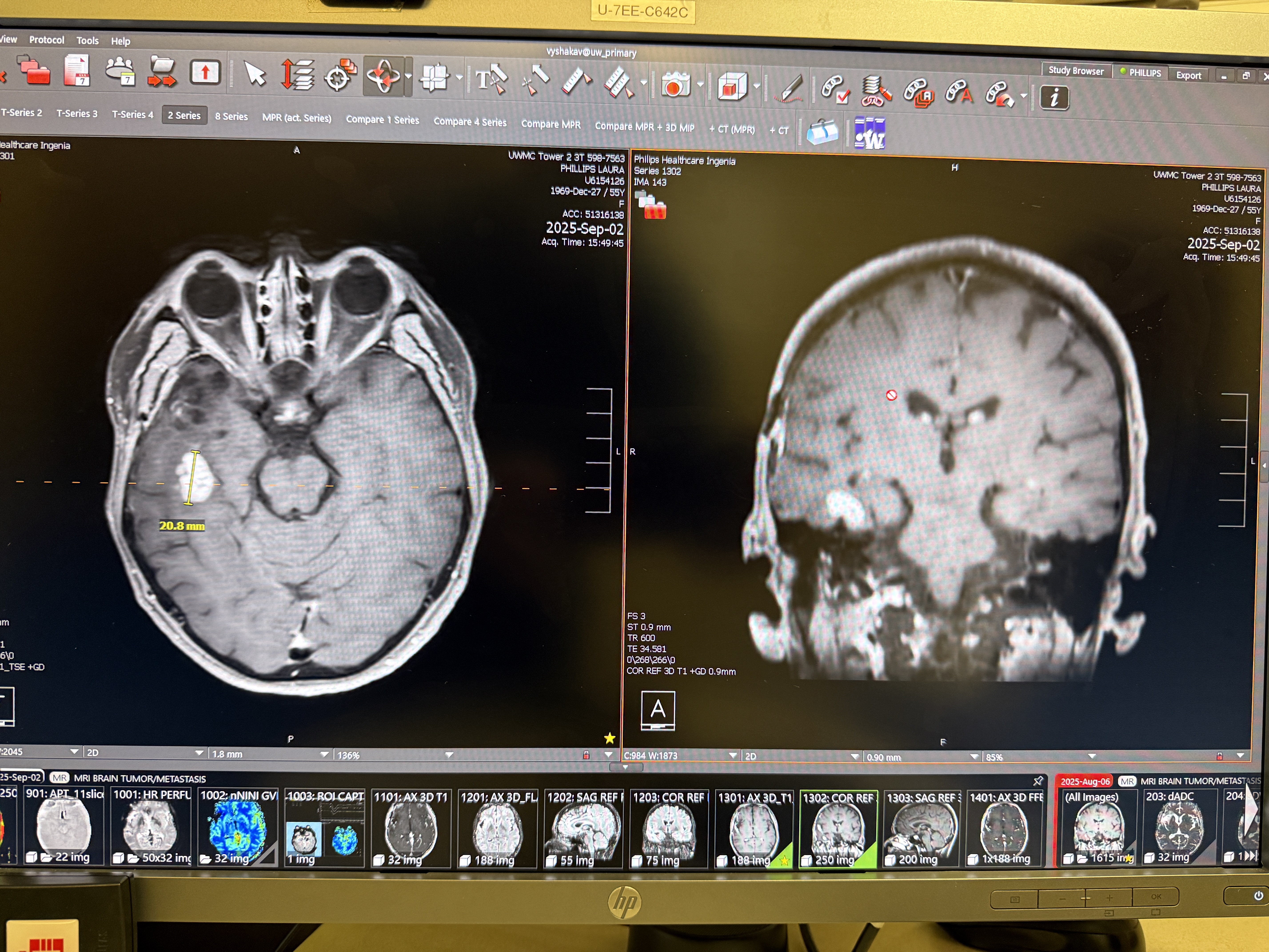
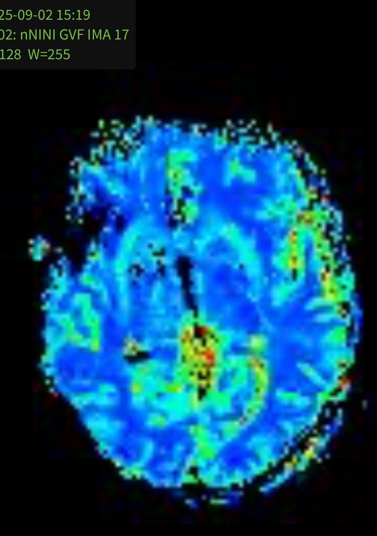
In the first image, you can see the spot on the left side of the image (right side of the brain since the images are reversed), but on the second image, you can see the area where the spot is does NOT show any highlights or additional blood flow.
What was also encouraging was that he said that even if it does turn out to be a new tumor, the location is easily accessible surgically and can be removed. We are hoping and expecting that is not the case.
We have a new MRI scheduled in a month, after which we will also be visiting our wonderful surgeon in Boise to have a check in.
As for our general health and state of being, we are both trying to spend more time exercising – mainly walking and taking advantage of the late summer sunshine here in the Seattle area and enjoying every day as we can.
What we are vigorously praying for is that highlighted spot will shrink and completely disappear and that it is not a tumor.
In the words, but not the spirit of Lady Macbeth: “Out, damned spot! Out, I say!”

Will continue to pray for complete healing.
LikeLiked by 1 person